Laparoscopic hysterectomy, often called keyhole uterus removal, is a groundbreaking surgical procedure that removes the uterus using minimally invasive techniques. This approach involves small incisions and specialized instruments, offering numerous advantages over traditional open surgery such as less pain, faster recovery, and reduced scarring. This article provides a detailed, step-by-step description of the laparoscopic hysterectomy procedure, from preoperative preparation to postoperative care and full recovery.
What is a Laparoscopic Hysterectomy?
A laparoscopic hysterectomy is a surgical method for removing the uterus through several small incisions in the abdomen using a laparoscope—a thin, lighted camera—and other surgical tools. Unlike traditional methods that require a large abdominal incision, laparoscopic surgery is less invasive, allowing for quicker healing, minimal blood loss, and less discomfort. This procedure is suitable for a variety of conditions including uterine fibroids, adenomyosis, endometriosis, abnormal bleeding, and gynecologic cancers in selected cases.
Preoperative Preparation

Consultation and Evaluation
Patients are generally instructed to stop certain medications, especially blood thinners, before surgery. Fasting is required from midnight the night before the procedure. Additionally, bowel preparation might be recommended to clear the intestines, ensuring optimal visibility and reducing infection risk during surgery. It is important to disclose any allergies and ongoing medical conditions to the surgical team.
Step-by-Step Laparoscopic Hysterectomy Procedure
Admission and Anesthesia
On the day of the surgery, you will be admitted to the hospital and prepared for the procedure. General anesthesia will be administered to keep you unconscious and pain-free throughout the operation
Surgical Positioning and Preparation
Once under anesthesia, you will be positioned on the operating table, typically in the lithotomy position, where you lie on your back with legs supported. The surgical team will cleanse your abdomen and pelvic area with antiseptic solutions to minimize infection risks. Sterile drapes are then placed to maintain a germ-free field
Creating Access – The Ports
The surgeon makes three to four small incisions, usually between 0.5 to 1 cm each, in the lower abdomen. One of these incisions will be at or near the navel for the laparoscope insertion, while the others serve as entry points for surgical instruments. These small “keyholes” create access points needed for visualization and manipulation during surgery.
Insufflation: Establishing the Working Space
Carbon dioxide gas is introduced into the abdominal cavity to gently inflate it. This creates a space between the abdominal wall and internal organs, providing the surgeon with a clear view and room to operate safely.
Insertion of the Laparoscope and Surgical Instruments
A long, slender laparoscope equipped with a camera is inserted through the navel incision and transmits high-definition images to video monitors in the operating room. Other instruments such as graspers, scissors, and energy devices are inserted through the additional incisions to perform the surgery.
Exploration and Assessment
Once all instruments are in place, the surgeon carefully inspects the uterus, ovaries, fallopian tubes, and surrounding structures to confirm the diagnosis and plan the specific steps needed during the operation.
Separation of Uterine Attachments
The uterus is carefully detached by sealing and cutting its supporting ligaments, blood vessels, and connective tissue with specialized energy devices. Structures such as ovarian ligaments (if ovaries are to be removed), fallopian tubes, round ligaments, and uterine arteries are sequentially divided with precise control to ensure minimal bleeding and injury.
Detachment from the Vagina
After mobilizing the uterus, it is separated from the top portion of the vagina, called the vaginal cuff. This delicate step requires caution to avoid injury to nearby organs such as the bladder and bowel.
Removal of the Uterus
Depending on the uterus size and surgical plan, it is removed either through the vagina or by morcellation, which involves dividing the uterus into smaller pieces to extract through the small abdominal incisions. Morcellation is avoided if malignancy is suspected.</p>
Inspection, Hemostasis, and Repair
The surgeon inspects the pelvic cavity thoroughly to confirm that bleeding has been controlled. The vaginal cuff is then sutured closed using laparoscopic suturing techniques or through the vaginal approach
Closure
Carbon dioxide gas is released from the abdomen, instruments are withdrawn, and the tiny incisions are closed with dissolvable stitches or surgical glue. Sterile dressings cover the wounds.
Transfer to Recovery
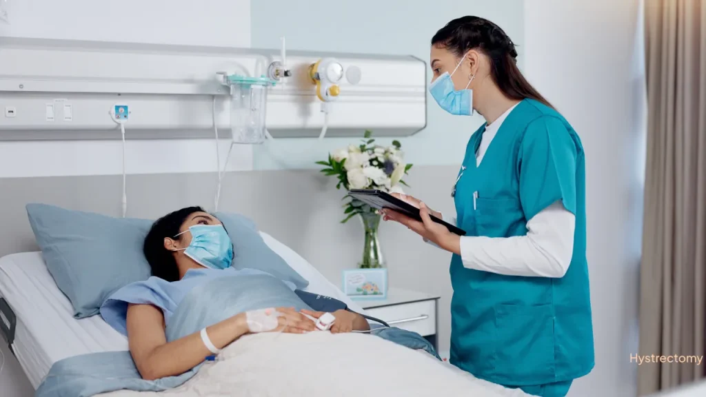
You will be moved to the recovery room, where healthcare staff will monitor your vital signs and overall condition as you wake from anesthesia. Most patients can expect hospital discharge within one or two days.
Immediate Postoperative Phase

Pain Management and Monitoring
Mild to moderate pain and cramping are common after surgery and can be managed effectively with oral pain medications. Nurses will watch for complications such as bleeding or infection during your hospital stay.
Mobilization
Early walking and movement are encouraged to prevent blood clots and promote normal bowel function.
Diet and Activity
Typically, patients start with small sips of fluids and progress to a regular diet as tolerated within the first 24 hours after surgery.
Hospital Stay
<p>Thanks to the minimally invasive technique, hospital stays for laparoscopic hysterectomy are generally brief, often just one or two days.
Full Recovery at Home
Wound Care
It is crucial to keep the small incision sites clean and dry. Bathing should be avoided in tubs or swimming pools until stitches are fully absorbed and wounds have healed, which usually takes two to three weeks.
Pain and Medication
Most patients find adequate pain control with prescribed oral medications. Should you experience severe pain, bleeding, or fever, seek immediate medical advice.</p>
Physical Activity
Light walking is encouraged soon after surgery, but heavy lifting, strenuous exercise, and sexual activity should be avoided for four to six weeks until your surgeon confirms it is safe to resume.
Follow-Up Visits
<p>A postoperative appointment is typically scheduled within one to two weeks to evaluate wound healing and address any concerns. Additional follow-up may be necessary depending on individual recovery.</p>
Menopausal Symptoms
If your ovaries were removed along with the uterus, you may experience menopausal symptoms such as hot flashes, mood swings, and vaginal dryness. Your doctor will discuss hormone replacemen t options if appropriate.
Advantages of Keyhole Uterus Removal
Laparoscopic hysterectomy offers many benefits compared to the traditional open abdominal approach. The smaller incisions result in less pain, reduced blood loss, minimal scarring, and a lower risk of infection. Patients benefit from quicker hospital discharge and faster return to normal activities. Additionally, the enhanced visualization provided by the laparoscope allows surgeons to operate with greater precision and safety.
Risks and Complications
While laparoscopic hysterectomy is generally safe, potential complications include bleeding requiring transfusion, injury to nearby organs such as the bladder or bowel, infection, formation of scar tissue, blood clots, and reactions to anesthesia. Choosing a skilled surgical team and modern equipment significantly reduces these risks.
